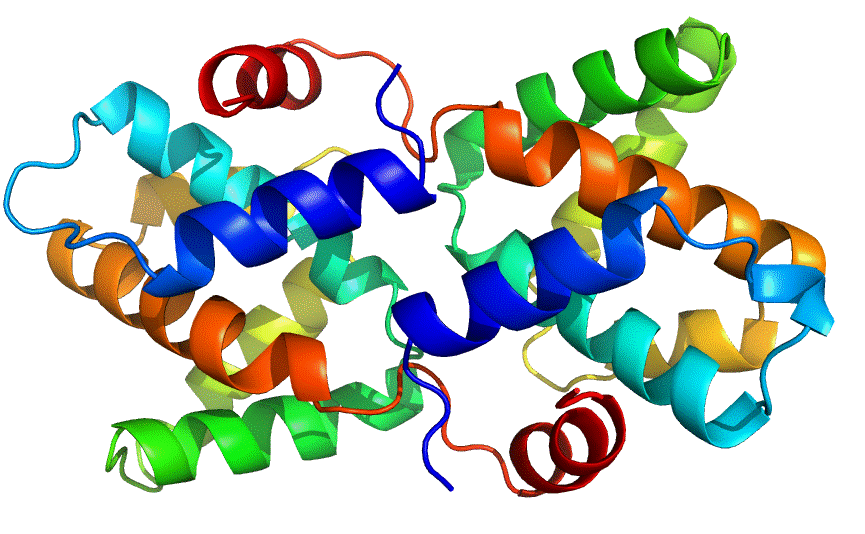
Ebolavirus VP30
The Ebola virus is the causative agent for Ebola hemorrhagic disease in humans and in other mammals. Since 1976 there have been approximately 2400 cases of Ebola reported; leading to the death of more than 75% of affected individuals. There are currently no known cures or vaccines for Ebola hemorrhagic disease. There are five known strains of Ebola, however only the Reston strain does not infect humans and only causes infections in other animals such as non-human primates and pigs. Understanding the differences between Ebola Reston and the more deadly strains of Ebola will be crucial to learning the mechanism of the disease. The genome of Ebola Reston is made up of seven genes, four of which are essential for viral transcription including the transcriptional cofactor VP30.
In collaboration with the laboratory of Erica Ollmann Saphire at the Scripps Research Institute in La Jolla, California; the Seattle Structural Genomics Center for Infectious Disease (SSGCID) has determined the second structure of the C-terminal domain of VP30 ever reported. The structure of VP30 from Ebola Reston closely resembles the structure of VP30 from the highly infectious Ebola Zaire. VP30 from Ebola Reston creates forms a homodimer (2 identical units of the protein complexed together); packing the C-terminal (end) alpha helix of one monomer into a conserved region on the face of the neighboring monomer. Superposition of VP30 Zaire and VP30 Reston shows that there is a significant rotation about the dimer interface, suggesting that the C-terminal domain of VP30 can adopt multiple conformations. The overall flexibility of the dimer interface may play a role in the modulation of RNA synthesis, the first stage of gene expression, or in another portion of the Ebola lifecycle.
This work was published in the Journal Acta Crystallographica Section F. Reference: Structure of the Reston ebolavirus VP30 C-terminal domain. Clifton M.C., Kirchdoerfer R.N., Atkins K., Abendroth J., Raymond A., Grice R., Barnes S., Moen S., Lorimer D., Edwards T.E., Myler P.J., Saphire E.O. Acta Crystallogr F Struct Biol Commun. 2014 Apr; 70(Pt 4):457-60. PMID: 24699737. Coordinates are available in the Protein Data Bank www.rcsb.org, PDBID 3V7O.
